Identifying rage cells in mice
Researcher Dayu Lin is working to identify the specific brain cells tied to aggression in mice. Dr. Lin and her colleagues have devised a method for manipulating that behavior by modulating the activity of the animal’s brain, culminating years of work exploring the neural circuits that regulate fighting in mice. Their findings are leading to a deeper understanding of the roots of animal aggression and possibly to novel medicines to quell violence in humans. Read the full article here: Illuminating the Roots of Violence.
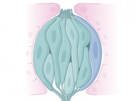
To locate rage at the cellular level, the researchers inserted electrodes into individual aggression cells in a mouse’s ventromedial hypothalamus (VMH) and monitored their activity. When the animal faced a foe, these cells chattered excitedly.
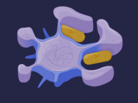
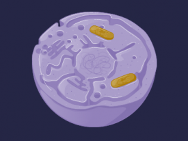
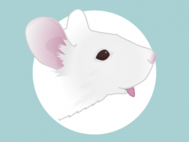
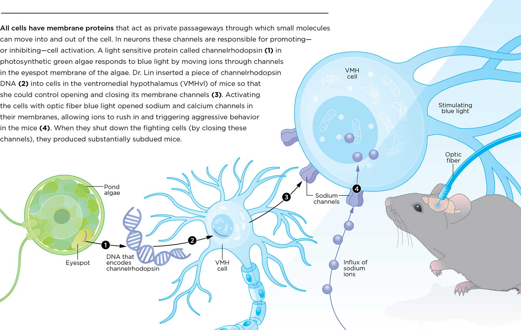
All cells have membrane proteins that act as private passageways through which small molecules can move into and out of the cell. In neurons these channels are responsible for promoting—or inhibiting—cell activation. A light sensitive protein called channelrhodopsin (1) in photosynthetic green algae responds to blue light by moving ions through channels in the eyespot membrane of the algae. Dr. Lin inserted a piece of channelrhodopsin DNA (2) into cells in the ventromedial hypothalamus (VMHvl) of mice so that she could control opening and closing its membrane channels (3). Activating the cells with optic fiber blue light opened sodium and calcium channels in their membranes, allowing ions to rush in and triggering aggressive behavior in the mice (4). When they shut down the fighting cells (by closing these channels), they produced substantially subdued mice.
Project: Infographic illustration and layout.
Client: NYU Physician

